- Special Feature
- Asbestos and Malignant Mesothelioma
- Published:
Pathology of mesothelioma
Environmental Health and Preventive Medicine volume 13, pages 60–64 (2008)
Abstract
The incidence of mesothelioma has been gradually increasing in Japan, and the underlying factor for this is considered to be the increase in the amount of asbestos imported into Japan between 1960 and 1975. Mesothelioma can be roughly divided into localized and diffuse types, but the former is extremely rare. In making a diagnosis of mesothelioma, it is important to confirm the location of tumor and the specific gross findings before histological examination. Mesothelioma can be categorized histologically as epithelioid type, sarcomatoid type, biphasic type, desmoplastic type, among others. It can take many forms; consequently, there are many diseases to be differentiated when the diagnosis of mesothelioma is based on histological analyses. Immunohistochemical stains are useful for making a diagnosis, but the correct combination of antibodies as positive or negative markers should be selected and a comprehensive assessment of the staining results is necessary. The accuracy of the pathological diagnosis is very important to the patients because they can be receive official compensation or relief when the diagnosis of mesothelioma is confirmed. Under present conditions, both clinicians and pathologists must make a concerted effort to improve the accuracy of the diagnosis of mesothelioma.
Incidence of mesothelioma
Mesothelioma is a malignant tumor that originates in the pleura, peritoneum, pericardium and tunica vaginalis, all locations where a lining of normal mesothelial cell is present. According to the official data of Ministry of Health, Labor and Welfare in Japan, the number of death due to mesothelioma was 500 in 1995, increasing gradually to 953 in 2004. This increase appears to parallel the increase of the amount of asbestos imported into Japan between 1960 and 1975, given that the latent period from the beginning of exposure to asbestos and an initial diagnosis of mesothelioma is approximately 40 years.
More detailed examination of the 878 patients who officially died from mesothelioma in 2003 reveals that the male:female ratio was almost 3:1 and that the peak of age of patients was 60+ in males and 70+ in females. The location of the mesothelioma was in the pleura in 84% of the cases, in the peritoneum in 12% and in the pericardium in 1%; there were not cases of mesothelioma in the tunica vaginalis. A larger proportion of females had mesothelioma in the peritoneum and pericardium than in the pleura [1].
General findings on mesothelioma
Mesothelioma can be roughly classified into localized and diffuse types, with the incidence of the latter being much higher than that of the former [2]. The localized form of mesothelioma was more frequently diagnosed in the past; however, most of the localized forms are currently diagnosed as a solitary fibrous tumor [3], which is a separate entity from mesothelioma based on immunohistochemical phenotyping (positive for CD34, a marker of primitive endothelial cell) and has no relation to asbestos exposure [4].
At the early stage of pleural mesothelioma, small nodules are found in the parietal pleura (not in the visceral pleura) that eventually extend along the pleural surface. Eventually, parietal and visceral pleurae show adhesion, and the tumor encloses the entire lung parenchyma. Very few cases of peritoneal mesothelioma have been reported at the early stage and, consequently, little is known in terms of pathology and disease progression during the early stage [5]. Most of peritoneal mesothelioma is found as a diffusely extensive tumor involving intestinal serosa or a large tumor located at the omentum or mesentery.
Following the initial diagnosis of mesothelioma, it is important to confirm the location of the tumor and its gross findings before histological examination. In the case of a pleural mesothelioma, a tumor in the lung parenchyma suggests lung cancer with a pleural extension. In females with peritoneal extension, the ovary should be carefully examined as the primary site of the tumor because the differential diagnosis between ovarian cancer and peritoneal mesothelioma is difficult and can only be made on the basis of histological analyses.
Histology of mesothelioma
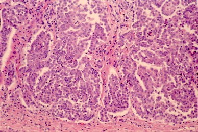
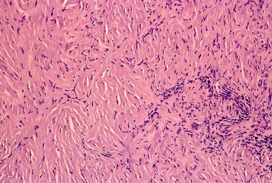
Histological classification of mesothelioma is shown in Table 1 [6]. There are three major types—epithelioid type, sarcomatoid type and biphasic type—and the proportion of each is approximately 60, 20 and 20%, respectively (Figs. 1, 2, 3) [7]. The desmoplastic type is rare (probably 1–2%) (Fig. 4), and special variants only appear sporadically (several percentages). However, the proportion of each histological type varies among the reports because of the large variety of histological analyses used [8–11].
Differential diagnosis
It is important to remember that there are many diseases to be differentiated when making a pathological diagnosis and that the tissue to be differentiated varies in terms of histological type (Table 2). Epithelioid types must be differentiated from lung adenocarcinoma for pleural mesothelioma [12], ovarian serous papillary adenocarcinoma or peritoneal serous carcinoma for peritoneal mesothelioma [13]. In terms of sarcomatoid types, sarcoma originating in the chest wall, lung, pleura, abdominal wall, peritoneum and intestine must be excluded. Sarcomatoid carcinoma (spindle cell sarcoma or pleomorphic carcinoma) of the lung is very difficult to differentiate from sarcomatoid pleural mesothelioma [14]. In terms of the biphasic type, carcinosarcoma or pulmonary blastoma of the lung, biphasic synovial sarcoma [15] of the pleural mesothelioma and carcinosarcoma of the female genital organs must be differentiated from their peritoneal counterpart. The desmoplastic type has a similar histology to fibrous pleuritis [16], and the differential diagnosis is very difficult, particularly if only a small biopsy specimen is available. Reactive mesothelial hyperplasia associated with pleuritis or other lung or pleural disease must be differentiated from early-stage epithelioid mesothelioma [17].
Usefulness of immunohistochemistry in an accurate diagnosis
The histochemical staining of hyarulonic acid and electron microscopic studies have been widely used in the past for making a differential diagnosis between mesothelioma and other tumors. However, immunohistochemical stains are currently the method of choice because of the simplicity and ease of these techniques. Many antibodies have been detected for use in immunohistochemical staining techniques aimed at diagnosing mesothelioma, but as yet there is no antibody that is completely specific for mesothelioma and on which a pathological diagnosis of mesothelioma can be singly based. Therefore, the combination of a number of antibodies as positive or negative markers is important, and an assessment of the results in a comprehensive manner is necessary (Table 3).
In the case of epithelioid mesothelioma, calretinin, WT1, thrombomodulin, mesothelin and D2-40 can be applied as a mesothelial cell marker [18–22]. CEA, TTF-1, Napsin A and surfactant apoprotein are used as markers for lung adenocarcinoma. In the case of ovarian serous papillary adenocarcinoma, we recommend CEA, Ber-EP4, MOC-31 and ER (estrogen receptor) as positive markers [23].
The antibodies chosen for sarcomatoid mesothelioma are very different from those used for epithelioid mesothelioma. In the case of sarcomatoid mesothelioma, cytokeratin (AE1/AE3 or CAM5.2 as antibodies) exhibits a high specificity and is the most useful [24]. On the other hand, because the diagnosis for true sarcoma is based on the specific differentiation of tumor cells, mesothelioma is eliminated by making its differentiation clear. For example, the following antibodies are known to be useful: MyoD1, desmin and myoglobin for rhabdomyosarcoma; desmin, α-SMA and h-caldesmon for leiomyosarcoma; S-100p for malignant nerve sheath tumor; KP-1 for malignant fibrous histiocytoma [25].
The most difficult tumor to be differentiated from sarcomatoid mesothelioma of pleura is sarcomatoid carcinoma (spindle cell carcinoma, pleomorphic carcinoma) of the lung. When immunohistochemical stainings are used, both respond positively to cytokeratin [24]. In this case, therefore, the gross finding or the clinical diagnosis by imaging is very important, as already mentioned.
Immunohistochemical stains may be useful in differentiating between fibrous pleuritis and desmoplastic mesothelioma. Desmin is positive for spindle cells of the fibrous pleuritis, while desmin is negative for tumor cells of the sarcomatoid mesothelioma [26]. The combination of EMA, desmin and p53 is useful for differentiating between reactive mesothelial hyperplasia and early-stage epithelioid mesothelioma. Reactive mesothelial cells are positive for desmin and negative for EMA and p53 [27].
Compensation or relief of patients
In the compensation system for occupational exposures to asbestos and in the new law for non-occupational exposure to asbestos, if the diagnosis of mesothelioma is certain, it can almost always be presumed to be related to asbestos exposure and the patients can receive compensation or relief. Therefore, the accuracy of the pathological diagnosis as mesothelioma is very important. However, a rough estimate is that approximately 10–15% of mesothelioma patients receive an inadequate diagnosis. In the committee for the judgement of patients’ relief, 30% of applicants are judged as not having mesothelioma or the decision is deferred until additional evidence is provided. It is therefore essential that Japanese clinicians and pathologists make an effort to improve the accuracy of the diagnosis as mesothelioma. In particular, the pathologists must improve the accuracy of the pathological diagnosis using adequate immunohistochemical stains.
References
Kishimoto T. Report of research on mesothelioma and occupational asbestos exposure in 2003 (in Japanese).
Allen TC, Cagle PT, Churg AM, Colby TV, Gibbs AR, Hammar SP, et al. Localized malignant mesothelioma. Am J Surg Pathol. 2005;29:866–73.
England DM, Hochholzer L, McCarthy MJ. Localized benign and malignant fibrous tumors of the pleura. A clinicopathologic review of 223 cases. Am J Surg Pathol. 1989;13:640–58.
Takeshima Y, Inai K. The pathological features of the pleural localized (solitary) fibrous tumors (in Japanese). Pathology and Clinical Medicine. 2004;22:708–12.
Nonaka D, Kusamura S, Baratti D. Diffuse malignant mesothelioma of the peritoneum: a clinicopathological study of 35 patients treated locoregionally at a single institution. Cancer. 2005;104:2181–8.
Travis WD, Colby TV, Corrin B, Shimosato Y, Brambilla E. (WHO). Histological typing of lung and pleural tumours. Berlin: Springer; 1999.
Attanoos RL, Gibbs AR. Pathology of malignant mesothelioma. Histopathology. 1997;30:403–18.
Shanks JH, Harris M, Banerjee SS, Eyden BP, Joglekar VM, Nicol A, et al. Mesotheliomas with deciduoid morphology: a morphologic spectrum and a variant not confined to young females. Am J Surg Pathol. 2000;24:285–94.
Galateau-Salle F, Vignaud JM, Burke L, Gibbs A, Brambilla E, Attanoos R, et al. Well-differentiated papillary mesothelioma of the pleura: a series of 24 cases. Am J Surg Pathol. 2004;28:534–40.
Yao DX, Shia J, Erlandson RA, Klimstra DS. Lymphohistiocytoid mesothelioma: a clinical, immunohistochemical and ultrastructual study of four cases and literature review. Ultrastruct Pathol. 2004;28:213–28.
Shia J, Qin J, Erlandson RA, King R, Illei P, Nobrega J, et al. Malignant mesothelioma with a pronounced myxoid stroma: a clinical and pathological evaluation of 19 cases. Virchows Arch. 2005;447:828–34.
Koss MN, Fleming M, Przygodzki RM. Adenocarcinoma simulating mesothelioma: a clinicophathological and immunohistochemical study of 29 cases. Ann Diagn Pathol. 1998;2:93–102.
Baker PM, Clement PB, Young RH. Malignant peritoneal mesothelioma in woman: a study of 75 cases with emphasis on their morphologic spectrum and differential diagnosis. Am J Clin Pathol. 2005;123:724–37.
Kobuke T, Yonehara S, Inai K, Tokuoka S. The diagnosis of sarcomatoid mesothelioma and the differential diagnosis between epithelioid mesothelioma and adenocarcinoma (in Japanese). Pathol Clin Med. 1987;5:1290–9.
Bequeret H, Galateau-Salle F, Guillou L, Chetaille B, Brambilla E, Vignaud JM, et al. Primary intrathoracic synovial sarcoma: a clinicopathologic study of 40 t (X;18)-positive cases from the French sarcoma group and the mesopath group. Am J Surg Pathol. 2005;29:339–46.
Cantin R, Al-Jabi M, McCaughey WTE. Desmoplastic diffuse mesothelioma. Am J Surg Pathol. 1982;6:215–22.
Churg A, Cagle PT, Roggli VL. Separation of benign and malignant mesothelial proliferations. In: Tumors of the serosal membranes. Silver Spring: ARP Press; 2006. p. 83–102.
Comin CE, Novelli L, Boddi V, Paglierani M, Dini S. Calretinin, thrombomodulin, CEA and CD15: a useful combination of immunohistochemical markers for differentiating pleural epithelial mesothelioma from peripheral pulmonary adenocarcinoma. Hum Pathol. 2001;32:529–36.
Hecht JL, Lee BH, Pinkus JL, Pinkus GS. The value of Wilms tumor susceptibility gene l in cytologic preparations as a marker for malignant mesothelioma. Cancer. 2002;96:105–9.
Miettinen M, Sarlomo-Rikala M. Expression of calretinin, thrombomodulin, keratin 5, and mesothelin in lung carcinomas of different types: an immunohistochemical analysis of 596 tumors in comparison with epithelioid mesotheliomas of the pleura. Am J Surg Pathol. 2003;27:150–8.
Kushitani K, Takeshima Y, Amatya VJ, Furonaka O, Sakatani A, Inai K. Immunohistochemical marker panels for distinguishing between epithelioid mesothelioma and lung adenocarcinoma. Pathol Int. 2007;57:190–9.
Ordonez NG. D2-40 and podoplanin are highly specific and sensitive immunohistochemical markers of epithelioid malignant mesothelioma. Hum Pathol. 2005;36:372–80.
Ordonez NG. Value of estrogen and progesterone receptor immunostaining in distinguishing between peritoneal mesotheliomas and serous carcinomas. Hum Pathol. 2005;36:1163–7.
Kushitani K, Takeshima Y, Amatya VJ, Furonaka O, Sakatani A, Inai K. Differential diagnosis of sarcomatoid mesothelioma from sarcoma and sarcomatoid carcinoma using immunohistochemistry. Pathol Int. 2008;58(2):75–83.
Lucas DR, Pass HI, Madan SK, Adsay NV, Wali A, Tabaczka P, et al. Sarcomatoid mesothelioma and its histologic mimics: a comparative immunohistochemical study. Histopathology. 2003;42:270–9.
Hurlimann J. Desmin and neural marker expression in mesothelial cells and mesotheliomas. Hum Pathol. 1994;25:753–7.
Attanoos RL, Griffin A, Gibbs AR. The use of immunohistochemistry in distinguishing reactive from neoplastic mesothelium. A novel use for desmin and comparative evaluation with epithelial membrane antigen, p53, platelet-derived growth factor-receptor, P-glycoprotein and Bcl-2. Histopathology. 2003;43:231–8.
Author information
Authors and Affiliations
Corresponding author
Rights and permissions
About this article
Cite this article
Inai, K. Pathology of mesothelioma. Environ Health Prev Med 13, 60–64 (2008). https://doi.org/10.1007/s12199-007-0017-6
Received:
Accepted:
Published:
Issue Date:
DOI: https://doi.org/10.1007/s12199-007-0017-6


